
Venous insufficiency skin manifestations/venous ulcers
R.C 18-05-23
- Diagnostic uncertainty.
- Suspected allergic contact dermatitis to bandages or topical medicaments or patients own topical agents – patch testing may be required.
- Non-healing venous ulcers (may need to biopsy in case SCC or pyoderma gangrenosum).
- Suspicion of large or small vessel vasculitis.
See https://apps.nhslothian.scot/refhelp/UlcerLegandFoot for comprehensive Foot & Leg Ulcer decision & referral pathway advice.
The combination of venous incompetence and insufficiency of calf muscle in promoting venous return, results in chronic venous hypertension, with stasis of blood in the veins of the legs. This may occur in the absence of visible varicose veins, though there are often small varicosities around the medial malleoli even if no varicose veins are visible higher up the leg.
Risk is increased with age, immobility, varicose veins and after deep vein thrombosis or cellulitis of lower leg.
Early diagnosis and management of venous insufficiency can reduce progression to irreversible changes and ulceration and the morbidity & cost associated with these.
Please see Vascular Surgery page on Refhelp re management of varicose veins. https://apps.nhslothian.scot/refhelp/VascularSurgery
Venous hypertension can result in several clinical presentations including:
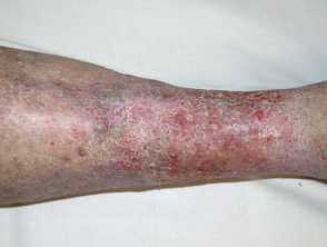
1. Venous eczema (VE)
Diagnostic features
VE presents as erythema +/- brown discolouration (due to haemosiderin deposits) in the gaiter area. Skin may be dry & itchy or weepy. Often bilateral (unlike cellulitis, which is also more likely to be tender and hot with an obvious border). Venous eczema affects approximately 20% of people over 70 years old. Legs may be oedematous with a history of gravitational oedema.
Management
1. Acute management
- Emollients – apply liberally and frequently (3-4 times a day if possible). Use ointments if very dry.
- Topical steroids – may help itch. Use moderate or potent topical steroid with steroid free periods. Severe or non- responding areas can be treated with a potent steroid as per LJF for up to 6-8 weeks, ideally under medicated bandages such as Viscopaste/Zipzoc.
- Topical calcineurin inhibitors – can be initiated by any health professional experienced in treating eczema, so can be used in Primary Care if GP comfortable to do so. Helpful if topical steroids required on regular basis. Switch steroid to tacrolimus ointment 0.1% which can be continued once daily for 6-8 weeks, reducing to maintenance therapy of 2 days per week. May cause burning sensation. Not usually used under occlusion.
- Infection – consider if patient pyrexial, or there is a unilateral area of tender, hot erythema. Antibiotics should be used as per LJF guidance but may be required for 10-14 days. If both legs look the same and the patient shows no evidence of infection, it is highly unlikely to be bilateral cellulitis.
- Pain can be a feature and can usually be managed with simple analgesia.
2. Long term management
- Treating the underlying venous insufficiency is key to preventing worsening eczema and the development of lipodermatosclerosis and venous ulceration.
- Patients should be prescribed compression hosiery (Class 2 if tolerated, Class 1 if not) after undergoing ABPI (“Doppler”) testing to ensure no arterial insufficiency. See Refhelp page https://apps.nhslothian.scot/refhelp/UlcerLegandFoot for guidance re acting on ABPI results.
- Encourage the patient to keep mobile & avoid prolonged periods of sitting if possible. Patients should elevate legs when resting.
- Support patients to lose weight if overweight/obese.
- Consider referral for venous surgery where superficial venous reflux (varicose veins) is present.
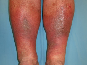
2. Lipodermatosclerosis
Diagnostic features
In the context of venous insufficiency and often associated with obesity, lipodermatosclerosis results from chronic inflammation & fibrosis, with leakage of fibrin from capillaries resulting in skin becoming hard and “woody” to touch. The gaiter area is red/brown in colour with small white areas of capillary atrophy (“atrophy blanche”). Fibrosis of the skin at the ankle results in swelling of the calves, giving the appearance of an “inverted champagne bottle” that can make bandaging challenging.
Management
As per venous eczema, though lipodermatosclerosis may require stronger analgesia as can be more painful than VE and may make compression more difficult to tolerate.
Potent or super potent topical steroids may be required to reduce inflammation in order to enable compression to be used, and the latter may need to be introduced gradually (e.g. Class 1 initially) to aid compliance.
Consider referral for venous surgery where superficial venous reflux (varicose veins) is present.
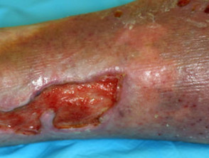
3. Venous ulceration
Please see Vascular Surgery & Ulcer Foot & Leg pages on Refhelp for information about other causes of leg ulcers.
Diagnostic features
These occur on the background of chronic venous insufficiency skin changes and account for 60-80% of leg ulcers.
They generally developing slowly over months and typically occur in the gaiter region of the leg (ankle to mid calf).
Varicose ulcers can be quite large but are relatively painless for their size (unless infected). Margins are generally irregular and the ulcer crater quite shallow (in contrast to the painful, smaller “punched out” ulcers caused by arterial disease) and there may be granulation tissue present.
It is important that patients are assessed for any co-morbidities that may compromise the healing of varicose ulcers (including poorly controlled diabetes/smoking and obesity).
1. Acute management
- Refer to health professional with experience in dressing venous ulcers (e.g. Practice Nurse or local CTAC service if available).
- Treat complications such as pain and infection (see eLJF for guidance).
- Start the strongest tolerated compression therapy if appropriate after ABPI assessment as soon as possible.
- SIGN Guideline 120 states that Pentoxifylline (400mg three times daily for up to 6 months) “should be considered” in patients with venous leg ulcers, but is unlicensed for this use and not on Lothian Joint Formulary.
2. Chronic management/prevention of ulcer recurrence
- As per venous eczema.
- Long term use of below knee graduated compression hosiery (strongest that is tolerated). Inform patients that this is likely to be needed indefinitely.
- Consider referral for venous surgery where superficial venous reflux (varicose veins) is present.
For Patients
https://www.bad.org.uk/shared/get-file.ashx?id=136&itemtype=document (venous eczema)
https://www.bad.org.uk/shared/get-file.ashx?id=237&itemtype=document (venous ulcers)
For Health Professionals
https://cks.nice.org.uk/topics/venous-eczema-lipodermatosclerosis/ (Venous eczema /Lipodermatosclerosis)
https://cks.nice.org.uk/topics/leg-ulcer-venous/ (Venous Ulcers)
https://www.formulary.nhs.scot/media/1430/wounds-dressing-selection-guide.pdf (Wound dressing)
https://www.pcds.org.uk/clinical-guidance/eczema-gravitational-eczema-syn-varicose-eczema-or-stasis-dermatitis (Gravitational eczema, Varicose Eczema and Stasis Dermatitis)