
BCC is the most common type of skin cancer (around 75% of all skin cancers diagnosed). It develops from basal cells, found in the deepest part of the outer layer of the skin (the epidermis).
They are typically found in areas of chronic sun exposure, such as the head and neck. It is rare for basal cell skin cancer to metastasise. However, they can cause significant destruction and disfigurement if neglected or inadequately treated.
Patients who have had a BCC are prone to developing further skin cancer (estimated clinical risk of 50% over 5 years).
Risk factors for all skin cancers include:
Excessive sunlight exposure and sun bed use (UV radiation) and is highest in people with fair skin colour and a susceptibility to burn.
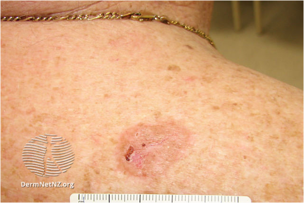
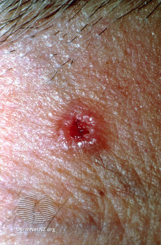
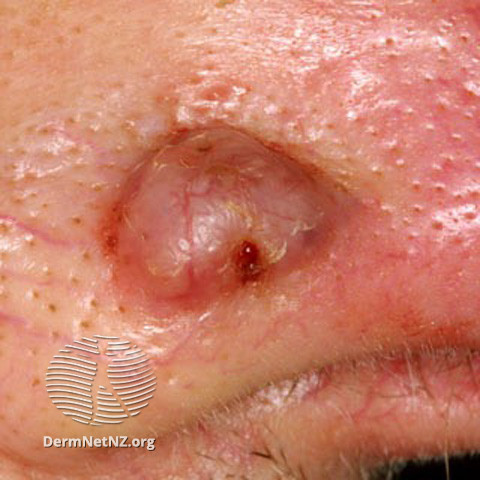
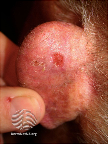
Suspect a BCC if a skin lesion has one or more of the following characteristics:
- Slow-growing lesion
- An ulcer with a raised rolled edge, a nodule on the skin (waxy or pearly), a reddish plaque, scar-like with tethering or contraction
- Prominent fine blood vessels within the lesion
- History of spontaneous bleeding
- May contain pigmented areas
- Itchy
- Rarely painful
All images on this page are sourced from DermNet | Dermatology Resource (dermnetnz.org)
Other skin lesions that are concerning for malignancy
The following skin changes should raise concern for a different malignant lesion (including Merkel’s cell carcinoma, sarcoma, or amelanotic melanoma):
- Nodule grows quickly (over weeks)
- A new change (growth, pigmentation, or pain) in a long-standing ulcer, scar, traumatic or inflamed area of skin
- Non-healing and/or destructive atypical ulcer
- Progressive unexplained scar-like area
- An unexplained skin lesion with loco-regional lymphadenopathy
Please refer these USOC via the Melanoma pathway.
B. C, C. L & P. O – 16-03-26
Refer high risk BCCs (those invading a potentially dangerous area e.g. eyelid margin, auditory meatus, nerve or major blood vessel of the face) USOC via Sci Gateway: Lauriston/SJH>Dermatology>LI Suspected High risk BCC.
Refer other BBCs on the head and neck urgently via Sci Gateway:
Refer BCCs on the trunk and limbs routinely via Sci Gateway: Lauriston/SJH>Dermatology>LI Dermatology Lesions.
Please use the Using the Consultant Connect App – RefHelp to take photos of the lesion(s) and then attach these to your Sci Gateway referral.
Low risk BCC can be removed in primary care where an appropriate service is available.
Criteria for removal in Primary Care (BCC):
- Send all specimens to pathology
- Diagnosis certain
- Well defined margins
- BCC should be excised with a 4 mm lateral margin and a cuff of fat in depth
For Patients
The BAD (British Association of Dermatologists) has a PIL on BCC. This can be found at: https://www.skinhealthinfo.org.uk/a-z-conditions-treatments/ & search for “basal cell carcinoma”.
For Healthcare Professionals
Primary Care Dermatology Society – Basal cell carcinoma
Scottish Referral Guidelines for suspected Cancer – Scottish Referral Guidelines for Suspected Cancer 2025
Dermnet nz – Basal Cell Carcinoma: Symptoms, Causes, and Treatment — DermNet