
The Scottish Cancer Referral Guidelines have been updated and went live on Wednesday, 6th August 2025. We are working hard to update all relevant information on the RefHelp website. If you would like to see the guidelines please click here Scottish Referral Guidelines for Suspected Cancer 2025 – gov.scot
Patients often present with new or changing occasionally symptomatic pigmented lesions.
It is normal for new naevi to develop until the age of 40 – referral should be based on suspicion of malignancy
Diagnostics Tips
- Malignant melanoma may be a new lesion or develop in a pre-existing mole
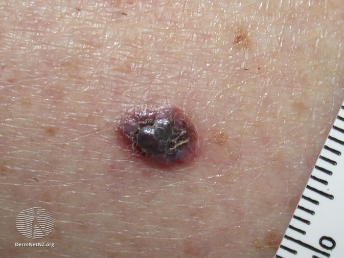
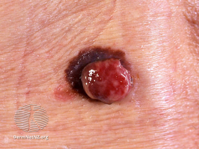
- Amelanotic malignant melanoma may present as an enlarging, granulating or smooth pink nodule. Beware the solitary pink papule!
- Assess risk factors:
- high sun exposure
- family history of melanoma
- presence of atypical naevi
- Changing naevus with suspicious ABCDE signs:
- Asymmetry
- Irregular Border
- Colour Variation
- Increasing Diameter
- Evolution-changing features
- Be suspicious of the “ugly duckling” mole that doesn’t look like its neighbours!
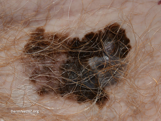
However these signs are not diagnostic and you should refer any lesion you feel may be malignant.
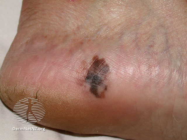
Acral-lentiginous melanoma
Melanocytic naevi and melanomas may occur under nails and on the palms of hands/soles of feet. Sub-ungual melanocytic lesions usually appear as a symmetrical pigmented band that starts at the base of the nail but does not involve the nail fold
The presence of pigment in the nail fold (“Hutchinson’s Sign”), ulceration or destruction of the nail plate or an asymmetrical/wide band are all highly suspicious for melanoma.
All images on this page are sourced from DermNet | Dermatology Resource (dermnetnz.org)
R.C 25-04-24
Suspected malignant melanoma
- Refer via “Urgent Suspicious Pigmented Lesion (Melanoma)” dermatology referral pathway and mark letter accordingly.
- If you have a high suspicion that the lesion may be a melanoma then tick the “high suspicion of melanoma” box.
- If it is a mole that you think is unlikely to be a melanoma but would like a specialist opinion on, then tick the “low suspicion of melanoma but wish to exclude diagnosis” box. Note that ALL referrals will be dealt with under the Scottish USOC pathway timeframe irrespective of whichever level of risk is chosen.
- Please use the Consultant Connect app to take photos of the lesion(s) and then attach these to your Sci Gateway referral.
- Attaching photos to referrals – RefHelp
Any patient who has a malignant melanoma removed in primary care must be referred urgently under the above pathway
- Provide education about moles e.g.British Association of Dermatologists Patient information leaflet (See Resources Tab)
- Give advice on sun protection & Vitamin D supplementation as per local guidelines.
- If any surgery performed, ensure the specimen is sent to pathology.
For Patients
The BAD ( British Association of Dermatologists) have several PILs on melanoma depending on the stage of the disease. These can be found at: https://www.skinhealthinfo.org.uk/a-z-conditions-treatments/ & search for “melanoma”.
For Healthcare Professionals
Primary Care Dermatology Society – http://www.pcds.org.uk/clinical-guidance/melanoma-an-overview1
Scottish Referral Guidelines for suspected Cancer
Dermnet nz – https://www.dermnetnz.org/topics/melanoma/